Crohn's disease is a chronic disease that causes inflammation in the digestive tract. While medications are often effective in treating and preventing symptoms, some people may require surgery if standard treatment does not work. One type of surgery that a doctor may recommend for people with Crohn's disease is bowel resection
. This procedure involves removing part of the small intestine.
In this article, we will discuss what a bowel resection is and what to expect before, during, and after surgery. We will also look at the risks and complications, prospects, and other types of surgery for Crohn's disease.
What is a bowel resection?
Bowel resection is a surgical procedure that doctors use to treat some people with Crohn's disease. When inflammation affects the intestines, it can sometimes cause a stricture. Strictures are areas of the intestine that become narrowed due to significant inflammation and block the passage of digested food. Unresolved strictures can lead to severe abdominal pain and cramps. There may be healthy tissue on either side of the intestinal stricture. During a bowel resection, the surgeon removes only the damaged part of the intestine and then connects the two healthy ends.
Preoperative testing
If you have symptoms of a bowel obstruction, your doctor will do a physical exam to check your stomach and bowel sounds.
Diagnostic evaluation may include an abdominal x-ray, computed tomography (ultrasound) scan, or ultrasound. These tests may or may not include a barium enema, which involves injecting a small amount of contrast material into the rectum to better visualize the structures.
If you have a blockage, you may also need a sigmoidoscopy or colonoscopy, which are invasive diagnostic procedures in which a camera is inserted into the colon to visualize the blockage.
When is bowel resection necessary?
People with Crohn's disease usually require surgery, according to the National Institute of Diabetes and Digestive and Kidney Diseases. Research shows that about 60% of people have had surgery within 20 years of being diagnosed with Crohn's disease. A doctor may recommend surgery for people with strictures that don't respond to standard treatment. Bowel resection may be necessary when other types of surgery, such as strictureplasty, are ineffective or not an appropriate option. A stricture can slow down the absorption of food and cause several symptoms, including:
- bloating;
- abdominal pain and cramps;
- nausea and vomiting;
- constipation.
These symptoms can be very serious and can lead to potentially dangerous complications, such as a perforation forming in the intestinal wall. The doctor may also recommend bowel resection to form fistulas. A fistula is an artificial channel that forms between two different parts of the intestine or connects the intestine to another organ. Fistulas can occur after severe inflammation of the intestinal wall. During a bowel resection, the surgeon will focus on removing the fistula and the damaged tissue around it.
Procedure parameters
There are several options for surgical treatment of intestinal obstruction. Surgery is usually urgent, which means you may have surgery within a few hours or a few days after you are diagnosed with a bowel obstruction. Some medications may help with nausea, but they do not prevent bowel obstruction from getting worse or better.
Ideally, you should not eat or drink for about eight to ten hours before this type of surgery, but due to the urgency, preoperative fasting is not always possible. Surgery for bowel obstruction is usually performed under general anesthesia in the operating room.
You may have open surgery with a large incision or minimally invasive surgery with several small incisions and a camera for imaging. It depends on the location, size and cause of the intestinal obstruction. Large tumors or widespread adhesions may require an open procedure, while a small tumor or infection may be treated with minimally invasive surgery.
How to prepare
Depending on the circumstances, the doctor may give certain recommendations to the person to prepare for surgery. These may include changes to the type or dosage of existing medications. Your doctor may also prescribe medications or antibiotics to prevent infection. A person may need to cleanse their intestinal tract the day before surgery. The doctor may advise the patient to use an enema, drink plenty of water, or drink a special solution that will help cleanse the intestines. Most surgeries require a person to fast from eating for a certain period before surgery. The doctor will tell the patient whether he needs to fast and for how long. Before fasting, it is best to eat healthy foods and avoid foods that can irritate your digestive tract.
Features of preparation for surgery
Before prescribing a resection, the doctor must conduct a visual examination and look at the medical history. The specialist will refer the patient for laboratory tests of urine and blood. Also, to confirm the need for surgical intervention, you need to obtain the result of an x-ray of the chest and abdomen.
If necessary, magnetic resonance imaging (MRI), electrocardiography (ECG), and computed tomography (CT) can be performed. Sometimes the patient is sent for laboratory tests aimed at assessing liver function.
Research results and a comprehensive diagnosis of the human body allow the doctor to identify problems in the patient’s intestines and prescribe a course of treatment.
Both the doctor and the patient should prepare for surgery. The patient must adhere to the following recommendations of the specialist: a week before the operation, it is forbidden to take medications (acetylsalicylic acid, drugs with anti-inflammatory and blood thinning effects).
You need to take antibiotics prescribed by the doctor 3-4 days before the procedure. You should also cleanse the intestines with an enema or laxatives, and a week before surgery you should start following a diet; it is advisable to exclude foods that contain fiber from your diet.
During the preoperative period, it is recommended to drink 2 liters of clean water during the day. 6-8 hours before the start of surgery, you are prohibited from eating and drinking.
During surgery
Before the operation begins, the anesthesiologist puts the patient under anesthesia. With general anesthesia, the person is unconscious and will not feel any pain during the procedure. There are 2 main types of small bowel resection: laparoscopic and open surgery. Laparoscopic surgery involves making small incisions in the abdomen to enter the abdominal cavity. The surgeon then inserts the laparoscope and small surgical instruments through the opening. A laparoscope is a thin tube with a camera and light at the end that allows the surgeon to see the abdomen using a monitor. During open surgery, the surgeon makes a larger incision and performs the procedure with standard surgical equipment.
After the surgeon has performed the bowel resection, he will stitch the incision and apply a bandage.
Types of operations on the small intestine
Surgical interventions on this organ are divided into two types: emergency and planned.
The following pathologies require emergency surgery:
- Meckel's diverticulum. Protrusion of the intestinal wall. The area is excised.
- Intestinal obstruction.
- Operation Whipple. Part of the pancreas, duodenum, gallbladder and lymph nodes is removed.
- Sticking. Adhesiolysis - elimination of intestinal adhesion is carried out in two ways: - laparoscopy - dissection with a laparoscope; - laparotomy - occurs through incisions on the abdominal wall.
In elective surgery, excision is indicated for tumors on the intestine.
Recovery after bowel resection
Full recovery after intestinal resection takes a long time, often up to 2 months. During this time, doctors regularly check the patient's condition. In general, doctors recommend avoiding activities that put stress on the abdomen, such as weightlifting or strenuous physical activity. They can also give the person additional exercise recommendations.
During recovery, the intestines must heal. A certain diet can help reduce stress on the intestines, promoting the healing process. Generally, recommendations include eating soft, easily digestible foods such as potatoes, rice and pasta.
Rehabilitation period
The rate of recovery after surgery depends on the volume of intervention performed and the general condition of the body. The rehabilitation period is also affected by the patient’s compliance with the medical and protective regime.
For the first day of the postoperative period, the patient is in the intensive care unit. Antibacterial, anti-inflammatory and analgesic drug therapy is carried out. Chemotherapy drugs may be administered according to indications.
Diet after resection:
- 12-24 hours after surgery, drinking water and unsweetened tea is allowed;
- It is necessary to expand the diet gradually. During the first week, the diet should consist only of liquid and pureed food;
- gradually you can include a small amount of solid foods in the menu;
- the patient will eat small portions 5-6 times a day;
- after a month, the patient can gradually return to his usual diet, but completely eliminate fatty, smoked, spicy and alcohol;
- with a properly selected diet there should be no digestive disorders.
To speed up rehabilitation, patients are recommended to engage in therapeutic physical education. The physiotherapist selects the volume and type of exercises individually for each patient.
Complications after bowel resection
Complications are possible after surgery. For example, a person may react inappropriately to an anesthetic. Infection and bleeding are also possible at the surgical site. In rare cases, the anastomosis may become disconnected. This complication is potentially life-threatening and requires immediate treatment. Other possible complications include kidney failure and fistula formation. The surgery may also lead to another complication called short bowel syndrome
. The small intestine is responsible for absorbing nutrients from food into the bloodstream. Removing too much intestine can lead to nutritional deficiencies in some people.
Indications
There are a number of pathologies for the treatment of which there is a need for this surgical intervention:
- traumatic injuries (in this case, emergency surgery is necessary);
- benign neoplasms that lead to partial or complete obstruction of the intestinal lumen or there is a high risk of malignant degeneration of the tumor;
- malignant neoplasms (if there is cancer, the operation is performed with regional lymphadenectomy to prevent relapse);
- complicated diverticulitis (diverticula are pouch-like protrusions of the intestinal wall that can appear in any part of the gastrointestinal tract, but most often occur in the large intestine);
- volvulus of the sigmoid colon, leading to intestinal obstruction;
- ulcerative lesions of the mucous membrane, which leads to severe discomfort for the patient (pain, constipation, frequent bleeding) and cannot be treated with medication.
Forecast
Bowel resection can help people experience relief from symptoms for many years. However, symptoms may eventually return. According to the Crohn's & Colitis Foundation, symptoms appear in about 50% of adults within 5 years of bowel resection. The inflammation usually affects the part of the intestine where the surgery was performed, but can occur in other places. Medicines can help treat these symptoms, but some people may need further surgery. Your doctor can advise possible treatment options if your symptoms return.
Principles of management of patients with short bowel syndrome
Short bowel syndrome is a complex of symptoms that develops after extensive resections of the small intestine. Similar operations are performed for Crohn's disease, intestinal ischemia, radiation enteritis, volvulus of the small intestine, desmoid tumors, trauma, etc. The prevalence of short bowel syndrome is difficult to assess, since its manifestations vary significantly in severity and not all cases are recorded by medical statistics. According to some estimates, in European countries, the prevalence of severe forms is 1.8–2 per 1 million population [5]. The length of the small intestine varies from 275 to 850 cm; in women it is shorter than in men. Under physiological conditions in healthy individuals, the total area of the actively functioning absorption and secreting surface of the small intestinal mucosa is colossal and comparable to the area of a tennis court (500 m2), and the area of the colon mucosa approximately corresponds to the surface area of a table tennis table (4 m2). A significant decrease in intestinal function due to resection leads to the development of specific short bowel syndrome and intestinal failure.
Short bowel syndrome is particularly severe in patients who have undergone resection with less than 2 m of small bowel remaining.
Based on the volume of resection, three main types of clinical and anatomical changes can be distinguished [8,13,16].
1. Resection of the small intestine while preserving at least part of the ileum, ileocecal valve and colon. Such patients are less susceptible to severe digestive disorders and rarely require long-term artificial nutrition.
2. Resection of the small intestine with the imposition of a jejunal-colic anastomosis. Patients with jejuno-colic anastomosis feel satisfactory for the first time after surgery, although signs of steatorrhea are noted. In subsequent months, trophological deficiency gradually manifests itself. However, over time, functional adaptation is possible with a decrease in the need for nutrients. Eating amounts of food that are normal for healthy people is accompanied by the appearance of diarrhea. If less than 50 cm of small bowel is retained, maintenance parenteral nutrition may be necessary.
3. Resection of the small intestine and colectomy with the application of a jejunostomy (jejunostomy). This option is characterized by diarrhea with the development of dehydration, electrolyte disorders (hypomagnesemia, hypocalcemia, hyponatremia) and trophological insufficiency in the near future after surgery. Diarrhea worsens after eating or drinking. Over time, no significant physiological adaptation of the intestine is observed. With jejunostomy, it is often necessary to constantly take isotonic solutions of sodium and glucose orally, take antidiarrheal drugs, and in some cases, parenteral nutrition and the administration of plasma substitutes.
The concept of intestinal failure. Intestinal failure syndrome (IF) involves disturbances in digestion and absorption, leading to the need for additional special nutrition and/or fluid and electrolyte support [11]. In the absence of adequate treatment, trophological insufficiency and/or dehydration develop in CI. The severity of CN can be most accurately determined by methods of calculating the energy balance that are inaccessible in practice. Based on treatment needs, CI is classified as mild, moderate, or severe.
• Mild: the need to select a special diet with a high content of nutrients and/or oral intake of glucose-saline solutions.
• Moderate: the need to prescribe special enteral nutrition and oral administration of glucose-saline solutions.
• Severe: the need for parenteral nutrition and administration of glucose-saline solutions.
As a rule, signs of CI develop when the length of the preserved segment of the small intestine is less than 2 m. Resection with preservation of less than 40–50 cm is considered to be the most unfavorable prognostically for the development of the most severe forms of CI.
CI can be acute or chronic, which is determined by the reasons for its development. Acute CI, as a rule, is a manifestation of infectious or radiation enteritis, intestinal ileus, and intestinal fistulas. Chronic CI develops as a result of surgical interventions (resection of the small intestine with the formation of anastomoses and stomas), and is also observed in Crohn's disease with damage to the small intestine, radiation enteritis, and gross motility disorders.
Pathophysiological changes after small bowel resection
Acceleration of passage through the intestines. During resection of the ileum and colon, due to disruption of the natural obturator mechanism, the rate of gastric emptying and passage through the small intestine increases significantly. This is mediated by low levels of production of YY-peptide and glucagon-like peptide-2, which are normally secreted by the cells of the mucous membrane of the ileum and colon and play an important role in the regulation of appetite and motility.
Loss of sodium and water. Under physiological conditions, passive secretion in the jejunum helps to achieve isotonic equilibrium between the intestinal contents and plasma. Most of the fluid must be reabsorbed in the jejunum, so significant losses of water and electrolytes may occur during resection and jejunostomy. If the length of the jejunum proximal to the stoma is less than 1 m, fluid loss through the stoma may exceed the amount ingested. If the patient ingests a hypotonic solution (with a sodium content of less than 90 mmol/l), additional loss of sodium occurs due to its diffusion from the plasma into the intestinal lumen along a concentration gradient.
Hypersecretion of gastric juice. In the first 2 weeks. after resection of the small intestine, hypersecretion of gastric juice is observed [20].
Impaired absorption and magnesium-calcium metabolism. After resection of more than 60–100 cm of the terminal portion of the ileum, malabsorption of fats, vitamin B12 and bile acids develops, which is not compensated by an increase in their synthesis in the liver. Unabsorbed bile acids enter the colon, where they stimulate the secretion of water and electrolytes. Unabsorbed fatty acids bind magnesium ions. At the same time, due to secondary hyperaldosteronism, the loss of magnesium in the urine increases. Hypomagnesemia is accompanied by a decrease in the activity of parathyroid hormone, inhibition of the production of D-1,25-dioxycholecalciferol, and calcium uptake in the renal tubules and intestines.
Adaptive processes after resection. With short bowel syndrome, compensatory hyperphagia often develops. In the intestine, there is an increase in the area of the absorbent surface - structural adaptation - as well as a slowdown in transit - functional adaptation.
In patients with jejuno-colic anastomosis, gastric emptying and passage through the small intestine may slow down over time due to increased activity of YY-peptide and glucagon-like peptide-2 in the blood; the intensity of absorption of nutrients, water, sodium and calcium gradually increases.
Patients with jejunostomy do not develop any significant adaptation [7].
Clinical assessment for short bowel syndrome consists, first of all, in characterizing the trophological status and water and electrolyte metabolism, including analysis of sodium, magnesium and calcium content.
Trophological insufficiency is manifested in a decrease in body weight by more than 10%, body mass index (BMI) <18.5 kg/m2, shoulder muscle circumference <19 cm in women and <22 cm in men.
With jejunostomy, assessing the degree of hydration, sodium and magnesium levels in the blood, monitoring body weight and the volume of discharge through the jejunostomy is extremely important. Signs of dehydration and sodium deficiency are usually thirst and arterial hypotension, up to the development of prerenal anuria. Severe sodium deficiency is characterized by a decrease in its concentration in urine <10 mmol/l.
For long-term prognosis and choice of treatment tactics, it is necessary to clarify the length of the remaining section of the intestine from the duodenojejunal ligament, which is possible during surgery or with contrast radiography.
A comparison of the features of the manifestations of short bowel syndrome with small-colic anastomosis and jejunostomy is presented in Table 1 [14].
General principles of patient management. The goal of treating patients with short bowel syndrome is to meet the body's water, electrolyte, and nutrient needs, while preferring oral/enteral nutrition over parenteral nutrition when possible. Management of patients by a team of specialists can prevent the rapid development of dehydration, reduce the risk of complications associated with CI and artificial nutrition (in particular, liver dysfunction, septic complications), and improve the quality of life of patients.
General principles for choosing the route of administration of fluids, electrolytes and nutrients in short bowel syndrome are presented in Table 2 [14]. In the most severe forms (jejunostomy with a portion of the remaining part of the jejunum less than 50 cm), it is vital to provide complete parenteral nutrition and rehydration. With a relatively long preserved segment of the small intestine and preservation of the passage through the large intestine (small-colon anastomosis), the diet and fluid intake approaches the natural one.
Features of management of patients with jejuno-colic anastomosis
Nutritional features. In the next 6 months. after surgery, given the tendency to gastric hypersecretion, antisecretory agents should be prescribed. When eating food by mouth, it should be taken into account that 50% of the energy or more may not be absorbed due to malabsorption, therefore the energy value of food should be increased; Feeding at night is indicated. If the effect is insufficient, parenteral nutrition should be added for several weeks or months [3].
The entry of long-chain fatty acids into the colon provokes an acceleration of transit, a decrease in water absorption, and thereby aggravates diarrhea. Fatty acids inhibit the activity of bacteria that ferment carbohydrates and bind calcium, zinc and magnesium. The latter contributes to an increase in diarrhea, absorption of oxalates and an increased risk of urolithiasis. When selecting a diet, preference should be given to the inclusion of medium- and short-chain triglycerides that can be absorbed in both the small and large intestines, for example, products based on coconut and palm oils, nutritional mixtures “Peptamen”, “Clinutren” [22]. Carbohydrates in the diet of such patients should be represented mainly by polysaccharides (in jelly, porridge and soups made from buckwheat, oatmeal, pearl barley, millet, corn grits, mashed potatoes, unsweetened and low-fat custard cream). Taking large amounts of mono- and oligosaccharides carries a risk of lactic acidosis as a result of overproduction of lactic acid by small intestinal lactobacilli and colon microflora at fecal pH above 6.5 [6].
With a preserved colon, significant disturbances in water and electrolyte balance are rarely observed. In case of a decrease in sodium content during the day, it is recommended to take an isotonic glucose-saline solution orally in an amount determined by the degree of dehydration (the usual physiological need is about 30 ml/kg of body weight).
In most cases, maintenance treatment with vitamin B12 and selenium preparations is necessary. In some patients, there is a need to compensate for the deficiency of zinc, essential fatty acids, and fat-soluble vitamins A, E, D, K.
Treatment of diarrhea. Diarrhea with short bowel syndrome is predominantly osmotic and partly hypersecretory and hyperkinetic in nature, caused by impaired secretion of gastrointestinal hormones. As the amount of food consumed by mouth is reduced, diarrhea improves, and in some cases temporary parenteral nutrition may be required. The principles of drug treatment of diarrhea are similar to those for jejunostomy: prescribing loperamide at a dose of 2–8 mg half an hour before meals, codeine phosphate at a dose of 30–60 mg before meals. If 100 cm or more of the terminal ileum is resected and bile acid malabsorption contributes to the development of diarrhea, it is advisable to prescribe cholestyramine.
In patients with small-colon anastomosis, episodes of confusion were recorded, the causes of which could be a decrease in magnesium content (<0.2 mmol/l), thiamine deficiency (Wernicke-Korsakoff syndrome, the main manifestations of which are severe memory impairment, coordination disorders in combination with polyneuropathy), lactic acidosis and hyperammonemia due to citrulline deficiency. Treatment consists of correcting magnesium deficiency, prescribing thiamine, an intermediate product of the urea synthesis cycle - arginine, and broad-spectrum antibiotics; in some cases, temporary parenteral nutrition is indicated [21].
Reducing the risk of cholelithiasis and urolithiasis. The incidence of calcium-bilirubin gallstones after intestinal surgery reaches 45%. The probable cause of cholelithiasis in these patients is a decrease in the activity (absence) of enterohepatic circulation of bilirubin and stasis of bile in the gallbladder and bile ducts. Prevention preferably includes enteral nutrition, administration of cholecystokinin, administration of ursodeoxycholic acid, and suppression of overgrowth of intestinal bacteria. Some surgeons prefer to perform prophylactic cholecystectomy when undergoing large-scale resection of the small intestine.
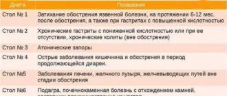
The risk of formation of oxalate urinary stones and nephrocalcinosis after small bowel resection reaches 25%. To prevent urolithiasis, one should prevent the development of dehydration, exclude foods rich in oxalates, and enrich the diet with medium-chain triglycerides and calcium (diet type No. 5).
Features of managing patients with jejunostomy. Unlike patients with small-to-colon anastomosis, with jejunostomy there is a significant loss of fluid and electrolytes, but there are no problems associated with severe bacterial fermentation in the colon.
When managing patients with a jejunostomy, it is necessary to monitor the volume of intestinal contents discharged through the stoma per day. A large volume of discharge may indicate the presence of unrecognized infectious complications, partial/transient intestinal obstruction, active enteritis (for example, clostridial).
Correction of water and electrolyte deficiency. Losses through the stoma increase after fluid and food intake, and each liter of discharge contains ≈100 mmol of sodium. If the length of the remaining jejunum is more than 50 cm, potassium loss is relatively small (≈15 mmol/L). Decreased potassium may occur due to hyperaldosteronism secondary to sodium loss; hypokalemia may also be a consequence of hypomagnesemia, leading to disruption of the transport systems that control potassium excretion [12]. With hypokalemia, there is a risk of heart rhythm disturbances.
A common mistake is the recommendation to take large volumes of hypotonic liquid orally to correct water and electrolyte metabolism; this leads to an increase in the volume of losses through the stoma. The same effect is observed when taking hypertonic glucose solutions, drinks with sweeteners, tea, coffee, alcohol, and fruit juices.
For patients with a jejunostomy, it is optimal to take isotonic rehydration solutions with a sodium concentration of ≈90 mmol/L (since the sodium concentration in the jejunostomy discharge is ≈100 mmol/L). The composition of rehydration solutions per 1 liter of plain water, recommended by WHO, includes: sodium chloride 60 mmol (3.5 g); sodium bicarbonate (or citrate) 30 mmol (2.5 g or 2.9 g, respectively); glucose 110 mmol (20 g). Alternative composition: sodium chloride 120 mmol (7 g), glucose 44 mmol (8 g). Examples of ready-made solutions for oral rehydration, generally corresponding to the composition recommended by WHO - Regidron, Oralit.
The volume of hypotonic fluid taken orally should not exceed 500 ml/day. It is recommended to eat solid and liquid foods separately (with an interval of at least half an hour).
If there is a large volume of discharge from the jejunostomy (more than 2 liters), it is advisable to completely exclude food and liquid intake for 2-3 days, which usually leads to a decrease in the volume of losses. An isotonic solution is administered intravenously in a volume of 2–4 l/day. The amount of daily urine should be at least 800 ml, and the sodium concentration in the urine should be at least 20 mmol/l. To maintain normal levels of potassium in the blood, correction of the balance of sodium and magnesium is of primary importance.
With a discharge volume of 1.5–2 l/day. It is recommended to drink a glucose-saline solution, and when taking a hypotonic liquid, add salt to the food (approximately 7 g, ≈2/3 teaspoon of table salt per day). If the volume of discharge is borderline (1–1.5 l/day), it can be recommended to drink regular liquid in a volume of less than 1 l and adding salt to food.
Adequate rehydration, which helps prevent the development of secondary hyperaldosteronism, is the most important step in the prevention and correction of hypomagnesemia. Magnesium salts are poorly absorbed from the gastrointestinal tract and can worsen diarrhea. When administered orally, preference is given to magnesium oxide in gelatin capsules at night, when intestinal transit is slowest. Excessive fat content in food should be avoided. If there is insufficient compensation for hypomagnesemia, 1-a-hydroxycholecalciferol is added in a gradually increasing dose (0.25–9.0 mcg/day). If it is necessary to prescribe magnesium sulfate parenterally, it is administered together with a solution of sodium chloride.
Correction of protein-energy deficiency during jejunostomy involves selecting a diet containing fats in the form of triglycerides and carbohydrates in the form of polysaccharides, which prevents an increase in food osmolarity. It is desirable that the concentration of sodium chloride in food is 90–120 mmol/l. The administration of an elemental diet (containing nutrients in an easily assimilated form - in the form of amino acids, oligopeptides, glucose, trace elements, dextrins, vitamins) has no advantages; on the contrary, it helps to increase the osmolarity of food and contains little sodium.
Medicines that suppress intestinal motility and secretion. If the above measures are insufficiently effective, they resort to medications that reduce the volume of discharge by inhibiting motility and/or secretion.
The treatment program for diarrhea in short bowel syndrome and intestinal stomas includes agonists of peripheral intestinal opioid receptors (loperamide, codeine phosphate, diphenoxylate). Drugs of this class slow down the passage of contents through the intestines by inhibiting propulsive contractions and stimulating segmental contractions. Accordingly, the degree of absorption of water and electrolytes increases. Opioid agonists also directly reduce the secretion of water and electrolytes and increase the tone of the ileocecal and anal sphincters.
Studies have shown that loperamide and codeine phosphate, by suppressing intestinal motility, reduce the volume of discharge from the ileostomy by 20–30% [14].
In one study, loperamide was administered at a dose of 4 mg 4 times a day. was more effective than codeine phosphate at a dose of 60 mg 4 times / day in reducing the volume of discharge through the stoma and sodium loss. The effect of co-administration of loperamide and codeine may be more pronounced.
According to British experts, the use of loperamide for short bowel syndrome is more preferable, since it does not have a central sedative effect and, as special studies have shown, does not cause fat malabsorption [14].
Loperamide has low systemic absorption when taken orally and does not penetrate the blood-brain barrier, so the risk of central side effects of the drug, including narcotic effects, is minimal.
As stated in the “Recommendations of the British Society of Gastroenterology” (2006), for short bowel syndrome, it is advisable to take the drug one hour before meals at a dose of 2–8 mg, the average dose is 16 mg/day. (in 4 doses). Given the frequent disruption of enterohepatic circulation of drugs after intestinal resection, it may be necessary to increase the dose of loperamide to 12–24 mg per single dose [14]. Increasing the dose must be carried out under the supervision of a doctor!
A number of randomized placebo-controlled studies have been conducted that convincingly prove the effectiveness and high safety of loperamide (Imodium) in the treatment of diarrhea in patients who have undergone intestinal surgery with various types of anastomoses and stomas [9,10,15,17,18].
The use of the combination of diphenoxylate and atropine is limited due to side effects associated with the anticholinergic effects of atropine.
In cases where tablets or capsules are released unchanged from the jejunostomy, their contents should be pre-mixed with food or water. For this category of patients, the ideal treatment option is lozenges - a unique dosage form of loperamide (Imodium). They instantly (in 2-3 seconds!) dissolve on the tongue and have a pleasant minty taste.
In cases of hypersecretion - when the volume of discharge exceeds 2-3 l/day. – the prescription of antisecretory agents is indicated: H2 blockers, proton pump inhibitors, octreotide, long-acting somatostatin/octreotide preparations. When the length of the jejunum is less than 50 cm, omeprazole should be administered intravenously.
If the ileum is preserved, mineralocorticoid preparations (fludrocortisone, aldosterone) or hydrocortisone can have a good effect in relieving diarrhea.
Alternative treatments. Data are accumulating on the effectiveness of the use of mucosal growth factors and hormone regulators (in particular, glucagon-like peptide-2) in stimulating adaptive processes after intestinal resection.
The introduction of short-chain fatty acids into the diet stimulates adaptation of the small intestine and significantly increases the absorption of amino acids [2]. Parenteral administration of glutamine and arginine, subcutaneous administration of growth hormone also accelerates the adaptation process [4,19].
Intestinal transplantation is a surgical treatment method that is indicated for patients with severe CI that cannot be corrected with conservative therapy (in particular, with a very short segment of jejunum - less than 50 cm, with severe motility disorders), as well as for patients with dangerous complications of parenteral nutrition (recurrent infection, progressive liver damage, etc.). Transplantation is also indicated when extensive bowel resection is necessary. More than 1,200 such operations have been performed worldwide. In some cases, a transplantation of the liver-small intestine complex and even a multivisceral transplantation (with the stomach and pancreas) is required.
Intestinal transplantation presents significant difficulties due to the high incidence of septic complications and low patient survival. Complications are caused by the temporary loss of the protective functions of the developed immune system of the intestinal mucosa and the presence of a non-sterile environment inside the lumen of the transplanted intestine. Immunosuppressive therapy includes tacrolimus-based regimens and antilymphocyte induction therapy. Today, intestinal transplantation is associated with 80% of patients surviving within a year, 50% of patients within 5 years, and in most cases without parenteral nutrition. The quality of life is comparable to that in uncomplicated CI.
Alternative surgical methods have been proposed to slow down peristalsis, in particular, reversion of a short segment of the small intestine, the essence of which is to “rotate” a section of the intestine that begins to contract in a retrograde direction.
Other types of surgery for Crohn's disease
Other surgical procedures are available to treat Crohn's disease. These include:
Stricturoplasty
For many people with a stricture, bowel resection may not be necessary. Instead, your doctor may recommend stricturoplasty, which is a type of surgery to widen the narrowed area without removing part of the intestine.
Colectomy
If Crohn's disease affects a long section of the large intestine, your doctor may recommend a colectomy. This procedure involves removing all or part of the colon.
Proctocolectomy
If inflammation affects an extended portion of both the large intestine and rectum, complete removal of both sections may be necessary. The surgeon will then connect the end of the small intestine to an opening in the lower abdomen to allow stool to be safely evacuated from the intestine.
When is bowel excision necessary?
Resection is a surgical intervention to remove the damaged part of the intestine with the formation of enteroanastomosis. The intestinal suture is applied using specialized devices or manually. At the clinic on Yauza, surgery on the small intestine is performed in extreme cases when therapeutic treatments are not able to cure the disease.
For what indications is resection necessary?
- extensive wounds resulting in injury to the abdominal cavity;
- ulcer;
- internal bleeding;
- peritoneal carcinomatosis;
- tumor-like neoplasms (benign and malignant);
- cancer;
- Crohn's disease;
- short intestine.
conclusions
For some people with Crohn's disease, your doctor may recommend a bowel resection to treat severe complications such as strictures and fistulas. The surgery involves removing the damaged portion of the small intestine and then reconnecting the healthy portions. Successful bowel resection surgery can allow a person to live for years without symptoms. However, in some people, symptoms may return and sometimes a second operation may be necessary. It is important for a person to take care of their body before surgery and during recovery to give themselves a chance to heal. Working closely with your doctor can help support proper recovery and reduce the risk of complications.
The authors of another study claim that patients with Crohn's disease are more likely to have anxiety disorders.
Advantages of performing surgery at GMS Hospital
GMS Hospital practices the most gentle methods of surgical treatment of intestinal diseases. We guarantee treatment as quickly as possible in the patient's situation. Our priority is minimally invasive interventions that shorten the rehabilitation period and minimize the risk of complications.
But this is not all the advantages of our surgery center. We provide:
- Competent medical care from highly specialized surgeons, as well as doctors of related specializations.
- A quick examination necessary for an accurate diagnosis. Diagnostics is carried out within 1 day.
- Effective treatment according to an individual program using innovative technologies. Including modern medical equipment: premium endoscopic units, vascular coagulators, new generation staplers, ultrasonic scissors, absorbable suture materials, and so on.
- Professional round-the-clock supervision by doctors and medical staff.
- Comfortable rooms with all amenities.
Surgery to remove intestinal cancer in the early stages
If the neoplasm is at an early stage of development and is small in size, the surgeon can completely remove it along with a segment of healthy tissue. This operation is called local resection.
The removed tissue is sent to a laboratory for further study. The pathologist will determine whether the tumor is high- or low-grade and whether a second, more extensive operation is necessary.
During the second surgery, more tissue that may contain cancer cells is resected, which reduces the likelihood of the disease returning.