The Khanty-Mansiysk Autonomous Okrug is located in the largest natural focus of biohelminthiases. Parasitic diseases such as opisthorchiasis and diphyllobothriasis are well known to the indigenous population of the Khanty-Mansi Autonomous Okrug. Every year, the district experiences population migration and an increase in these diseases in the district. For an uninformed person, avoiding infection with these diseases is very problematic.
Opisthorchiasis and diphyllobothriasis are helminthic diseases of humans and animals, carried by river fish.
How can you become infected with these diseases?
- When eating raw (patanka, stroganina), insufficiently processed, lightly salted and dried fish.
- When using non-neutralized equipment after cutting fish (knives, dishes, equipment) for preparing ready-made dishes (salads, cold appetizers).
- If personal hygiene rules are not observed while cutting raw fish or after finishing it (poorly washed hands);
- When taking a sample during the preparation of fish dishes.
Opisthorchiasis
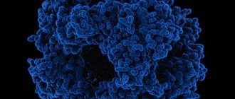
Opisthorchiasis is a natural focal, severe helminthic disease of humans and carnivores, with primary damage to the liver, bile ducts, and pancreas. The causative agent is the Siberian or cat fluke (opisthorchis felineus). The parasite is small, 8-13 mm, oval in shape with two suckers. In the human body, the number of parasites can reach up to 40 thousand copies.
The lifespan of the parasite in the human body is up to 25 years. The cat fluke (opisthorchid) requires three hosts to develop.
The definitive host is humans, domestic animals (dogs, pigs, cats), wild animals (foxes, otters, arctic foxes) and other species of mammals whose diet includes fish. Eggs that are not dangerous to humans and animals are released from the intestines of the definitive hosts into the environment. Once in a pond, the eggs can remain viable for 5-6 months.
The intermediate host is mollusks. The eggs are swallowed by mollusks and, together with food, enter the gastrointestinal tract of the mollusk, where they turn into larvae. Mollusks hatch into thousands of larvae that float freely in the water, but are not dangerous to humans, so you cannot become infected with opisthorchiasis through water.
An additional host is carp fish. In fish, the larvae are localized in the muscles, eyes and other organs. When consuming fish infected with opisthorchid larvae, a person or animal becomes the definitive host.
Once in the body of the final host, the larvae, under the influence of gastric juice, are freed from the capsule shells, exit into the intestines and penetrate the liver, gallbladder, and pancreas, causing a severe disease. The accumulation of parasites in the bile ducts contributes to their blockage, disruption of the outflow of bile and deformation of the duct.
The disease can occur in acute or chronic form.
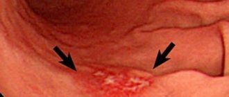
The acute form of the disease is caused by severe intoxication of the body and is characterized by increased body temperature, headache, malaise, swelling, rash, pain in muscles and joints. These symptoms occur 18-45 days after infection. In severe cases, the development of allergic myocarditis and eosinophilic small focal pneumonia is possible.
The chronic form of opisthorchiasis is characterized by symptoms of inflammation of the gallbladder (cholecystitis) with periodic exacerbations and remissions. Patients are concerned about heaviness and periodic pain in the right hypochondrium, epigastrium, loss of appetite, nausea, bloating, constipation or loose stools with the development of dysbacteriosis.
Complications
If treatment is not started on time, the disease becomes chronic and leads to consequences. Parasites feed on the mucus of internal organs and blood. Also, the life process of the larvae cannot do without their decay, which poisons the body with toxins.
The consequences of opisthorchiasis are very serious:
- liver damage by types of cirrhosis;
- abscesses may occur on the liver;
- the occurrence of peritonitis or pancreatitis;
- the gallbladder may become inflamed;
- the pancreas becomes inflamed;
- liver cancer;
- pancreas cancer;
- anemia;
- chronic hepatitis;
- development of asthma;
- general allergic reactions of the whole body;
- heart problems;
- risk of gastritis and stomach ulcers;
- epileptic seizures.
Diphyllobothriasis
Diphyllobothriasis is a helminthic disease affecting the digestive organs. The causative agent is the broad tapeworm. This is the largest of the human helminths, its length can reach 10 and sometimes 20 meters. The parasite consists of a head, neck and body. The head is oblong oval in shape, flattened on the sides and has on its narrow sides two longitudinal suction slits (bothria), with which the tapeworm is attached to the intestinal wall. The body consists of many segments, and the width is much greater than the length, which is due to the name of the parasite (wide tapeworm). The number of segments can reach 3000-4000 pieces. The tapeworm lives in the upper parts of the small intestines, feeds on the entire surface of the body, while absorbing various nutrients, including vitamins Bi2 and folic acid. The wide tapeworm is hermaphrodite. During the day, up to 2 million eggs are released into the external environment with feces. The number of parasites can reach up to 100 copies. The lifespan of parasites in the human body reaches 28 years.
For the development of a wide tapeworm, like opisthorchiasis, the presence of three hosts is necessary.
The definitive host is humans, domestic and wild animals. All of them secrete eggs, which end up in water bodies with meltwater. Diphyllobothriasis cannot be contracted through water.
The intermediate host is Cyclops (crustaceans). The eggs are swallowed by crustaceans (cyclops) and larvae develop in their bodies. Cyclops are swallowed as food by freshwater predatory fish.
Additional hosts are fish of predatory species: pike, burbot, perch, ruff; pike caviar is especially dangerous.
Having attached themselves to the wall of the intestines, the parasites injure the intestinal mucosa with bothria and can be one of the reasons for its necrosis. Sometimes intestinal blockage occurs.
Diphyllobothriasis occurs in mild or severe form, which is associated with the intensity of invasion, the presence of concomitant diseases and the general condition of the body. Sometimes the disease is asymptomatic.
In mild cases, patients complain of general weakness, poor appetite, nausea, pain and rumbling in the abdomen, intestinal disorders, and decreased ability to work.
In severe cases, intestinal obstruction occurs. In 2-3% of patients, a severe form of anemia (anemia) occurs. Patients complain of weakness, drowsiness, dizziness. Bright red spots and cracks appear on the tongue. The skin becomes pale with a yellowish tint; The liver and spleen may enlarge. Body temperature reaches 36-38 degrees.
The diagnosis of these diseases is established based on the detection of tapeworm and opisthorchid eggs in the feces.
Diagnostics
Photo: parazity.com
When diagnosing opisthorchiasis, it is important to clarify with the patient whether he is engaged in fishing, whether he has recently traveled to river areas, whether he has eaten poorly processed or raw fish, and the presence of the disease in people with whom the patient is in contact. A general examination can reveal a rash in typical places (upper torso, upper limbs), jaundice of the skin and icteric sclera, enlarged liver and pain on palpation in the right hypochondrium and epigastrium.
Laboratory diagnosis consists of detecting in a general blood test an increase in leukocytes, eosinophilia (a marker of allergic reactions and helminth infestation), and an increase in the erythrocyte sedimentation rate. In the biochemical blood test, there is a clear increase in liver enzymes (ALT, AST, ALP, GGT) and bilirubin.
Serological tests play an important role. Using an enzyme immunoassay using opisthorchiasis antigen, anti-opisthorchiasis antibodies of classes G and M are detected in the blood serum of patients. In some cases, false-positive test results are possible if the patient has other parasitic diseases (echinococcosis, diphyllobothriasis, ascariasis, etc.)
Special research methods to exclude helminthic infestation are called helmintho-ovoscopic. The essence of the technique is to search for worm eggs in separated liquids. Directly for opisthorchiasis, it is important to search for eggs under a microscope using special dyes in feces and duodenal contents, which are collected during FGDS.
To clarify the diagnosis, instrumental diagnostic methods are prescribed. Fibrogastroduodenoscopy shows the functional state of the stomach, duodenal bulb and sphincter of Oddi (the place in the lumen of the duodenum where the common bile duct exits)
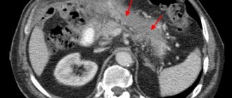
Ultrasound examination is used to clarify the condition and presence of inflammation of the bile ducts.
If the clinical picture is unclear, X-rays, MRIs, and CT scans of the abdominal organs may be required.
Prevention of helminthic diseases
- cook the fish for 15-25 minutes;
- cooking cutlets, meatballs, etc. - 15-25 minutes;
- baking pies with fish for at least 45-60 minutes;
- cold smoking of fish must be carried out either after pre-salting for 2-3 days, or after freezing;
- fry large pieces of fish spread out and always in fat for at least 20 minutes, fry small fish whole for 15-20 minutes;
- salt the caviar (at a temperature of 5-6°C), the ratio of the amount of salt is 6% to the weight of the caviar for 12 hours, for example: 60 g of salt per 1 kg of caviar, or salting the caviar in a 5% solution (50 g of salt per 1 kg of caviar ) at least 2 days with periodic mixing of caviar;
- salt: small fish for 14 days, large fish (over 25 cm) for 40 days with the addition of 2 kg of salt per 10 kg of fish;
- drying of fish for at least 3 weeks with preliminary salting for 2-3 days; d freezing: at 40°C-7 hours, at 35°C-14 hours, -28°C-32 hours.
The danger of contracting opisthorchiasis and diphyllobothriasis does not disappear all year round.
Why should you be careful with sushi?
Japanese cuisine is gaining more and more popularity, and the number of restaurants that offer sushi and rolls is growing every year. Therefore, many people are interested in whether sushi is dangerous for health? With all the obvious benefits of sushi, they are also fraught with danger. To prepare Japanese cuisine, raw fish is used, which means it may contain opisthorchid larvae.
Parasites do not die even when frozen and can live for up to a week. Since in standard freezers the temperature is not lower than -18 degrees, and to destroy the larvae requires freezing to -40 degrees. Therefore, sushi can cause opisthorchiasis.
Medicines
Photo: expertdent.net
To eliminate the allergic reaction, drugs from the group of H1-histamine receptor blockers (Loratadine, Cytherizine, Clemastine, Chloroperamine) are used.
To eliminate spasm of the biliary tract and the associated pain syndrome, antispasmodic (Drotaverine, Papaverine, Metamizole, Mebeverine) and choleretic drugs (Ursodeoxycholic acid, Ademethionine) are used. Such drugs are prescribed from the beginning of treatment and used for at least three months.
If indicated, it is possible to prescribe drugs that enhance intestinal motility (Metoclopramide, Domperidone), as well as pancreatic enzymes (Creon, Pancreatin), probiotic drugs (Linex, Bifiform, Hilak) and drugs for eradication therapy.
For chemotherapy, the anthelmintic drug Proziquantel is used. The drug has a wide spectrum of action and is used not only for opisthorchiasis, but also for other helminthic infestations. Affects the muscles of parasites, causing paralysis. The course of treatment is 2 days, during which the patient takes 6 doses of the drug. Contraindications for use are pregnancy and acute complications of opisthorchiasis.
During the rehabilitation period, probiotics, hepatoprotectors (Phospholipids, Thioctic acid, Ademethionine), and mineral medicinal table waters are prescribed. The rehabilitation period is aimed at the complete restoration of the body.
Methods for determining infestation
To detect opisthorchiasis in fish, fishing enterprises use several diagnostic methods simultaneously. To do this, several samples are caught, at different stages of development, if possible, and if the reservoir is large, in different places. Which fish is infected and whether there is opisthorchiasis in the eggs can only be found out through clinical trials.
Types of laboratory tests:
- Physico-chemical, detect deviations in the interaction of ammonia released from decomposed fish and hydrochloric acid.
- Sanitary and microbiological, fish muscle tissue is checked by applying rosolic acid, coloring healthy areas in shades of pink. Uncolored areas indicate the presence of infection.
- Parasitological, muscle fibers and subcutaneous tissues are checked under a microscope. Only with the help of special equipment can you see what opisthorchiasis looks like, which fish contain opisthorchiasis, and record them in a photo.
- Organoleptic, a rarely used type of research, is carried out only when there is a significant deviation in the development of fish.
What fish are infected with this helminthiasis?
The cat fluke chooses as its host fish living in fresh open water bodies; most often, representatives of cyprinids are infected. Sea and ocean inhabitants do not suffer from this helminthiasis, since they live in salt water. Red fish can only become infected if they are bred in fresh water bodies.
In which fish is opisthorchiasis most often detected?
Among the inhabitants of open fresh water bodies, opisthorchiasis affects representatives who feed on infected mollusks, crustaceans or small fish.
Most often infected with opisthorchiasis are ide, perch, carp, dace, ram; much less commonly, the mentioned fish parasites affect sterlet.
The list of fish carriers of opisthorchiasis also includes the following:
- bream, carp, roach, carp, crucian carp (representatives of the carp family);
- perch, ruff, catfish, wrasse (perch);
- thorn, sturgeon (sturgeon);
- Karelian trout, musk, nelma, white fish, grayling, peled or cheese (salmon).
- Many representatives of the salmon family are considered delicacy varieties, which are present in raw form in most dishes, and although salmon rarely get helminthiasis, there is still a risk of infection.
- Trout belongs to the salmon family, so it is practically not susceptible to helminthiasis, but Karelian trout, which is grown in artificial reservoirs, is easily infected with cat fluke.
- Previously, it was believed that sterlet and sturgeon do not suffer from helminthiasis, but recent data indicate that some of them also contain parasite larvae due to contamination of water bodies with waste (feces, sewage, garbage).