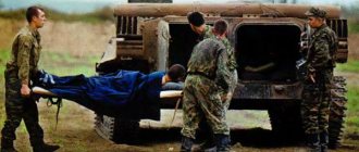
Assisting with acute gastrointestinal bleeding
Urgent Care:
- strict stretcher regime;
- in case of collapse - transportation in the Trendelenburg position;
- Eating food and water is prohibited;
- “coldness” (if any) on the stomach;
- infusion of plasma-substituting solutions: dextran/sodium chloride at the rate of 10 ml/kg body weight, 10% hydroxyethyl starch solution, then - when blood pressure is more than 80 mm Hg. Art. - drip;
- administer sodium ethamsylate 2-4 ml of 12.5% solution intravenously;
- oxygen therapy;
- emergency hospitalization in the surgical department.
Clinical manifestations
The acute form is characterized by severe girdling pain radiating to the left scapula and collarbone, vomiting that does not bring relief, dizziness, tachycardia, weakness, and stool disturbances. The temperature depends on the stage of pancreatitis. If in the first two days it is normal (with the exception of acute cholangiopancreatitis and cholecystopancreatitis), then from the third it increases. Characteristic symptoms of the disease are bluish spots throughout the body. The reason is circulation problems.
There are four stages of acute pancreatitis: enzymatic, reactive, period of purulent complications and outcome.
Enzymatic (early) lasts the first five days. Characterized by severe symptoms of intoxication.
Reactive begins on the 7th day of the disease and lasts 7-10 days. Manifested by fever and gastric symptoms.
Period of purulent complications. Foci of necrosis of the gland become infected and the bacterial process spreads to neighboring organs. Possible bleeding.
Outcome: for a mild form, recovery from inflammation takes 3 weeks, for a severe form it takes from a month to two.
Medicines for pancreatitis are prescribed exclusively by a doctor. Self-medication can be fatal.
Providing assistance with a strangulated hernia
Urgent Care:
- emergency hospitalization in the surgical department, transportation on a stretcher;
- it is strictly forbidden to reduce the hernia, use analgesics, baths, or heat;
- Hospitalization is also indicated if by the time of examination the strangulated hernia has been reduced;
- patients with an irreducible hernia and pain syndrome should also be hospitalized in a hospital for emergency reasons.
previous article
Vitamin D and children's health
next article
Main differential indicators in children
Up
What can cause the disease
Among the provoking factors, the following should be noted: excessive alcohol consumption, inflammatory diseases of the duodenum, hormonal disorders, trauma (wounds) of the abdomen, side effects of certain medications (antibiotics, estrogens, antitumor drugs) and pathologies of the pancreatic duct. Vascular diseases, helminthic infestations, metabolic disorders and surgery on the biliary tract can also become a “trigger” for the development of the disease. Read on to learn more about the symptoms of pancreatitis in adults.
Diagnosis and treatment of acute pancreatitis (AP) is the task of surgeons. However, the variety of its clinical manifestations, especially in cases of severe, complicated course, does not exclude the situation when the patient is admitted to a therapeutic hospital in critical condition or under the guise of another disease. This is why it is useful to be aware of such conditions, be able to recognize them and provide the necessary assistance before the consultant surgeon arrives.
For a clearer understanding of the complications and features of the course of AP, we should briefly dwell on the main stages of its pathogenesis (Fig. 1).
Figure 1. Scheme of the typical course of TOP by phases and periods with corresponding characteristic complications.
As the course progresses, severe AP (SAP) goes through phases of pancreatogenic toxemia and destructive complications. Within the phases, periods are distinguished that have different pathogenesis, typical complications and clinical features inherent in them. For the period of acute hemodynamic disorders, which can last from the first few hours of the disease to 2-3 days, the leading moment of pathogenesis is the release of cytokines and inflammatory mediators with the development of systemic respiratory and circulatory disorders up to pancreatogenic shock and adult respiratory distress syndrome (ARDS). Typical local complications of a destructive nature are local edema, liquid impregnation of the retroperitoneal tissue in areas of pancreatogenic destruction and free liquid in the serous cavities. For the period of multiple organ failure, which can last from the first 2-3 to 10-12 days of the disease, the leading point of pathogenesis is reperfusion of areas of necrosis and necrobiosis, reperfusion syndrome and resorptive toxemia up to the development of early multiple organ failure. Typical local destructive complications of this period are acute fluid accumulations in the omental bursa and retroperitoneal tissue. The period of aseptic destruction, which, as a rule, occurs in the 2nd week of the disease, is characterized by a pancreatogenic infiltrate, which is an array of sterile retroperitoneonecrosis with an infiltrative-inflammatory shaft surrounding it, followed by resorption, sequestration or infection (Fig. 2).
Figure 2. Scheme of the structure of pancreatogenic infiltrate at the beginning of the phase of destructive complications and typical options for its further evolution. When infected, purulent complications of a limited nature develop (pancreatogenic abscess or early infected pseudocyst) or unrestricted retroperitoneal pancreatogenic phlegmon. The indicated schematic classical course of TOP with a sequential change of periods and phases does not always occur. In practice, we often have to deal with a situation where the complications of the previous phase that have not yet been resolved are superimposed by the clinical manifestations of the next one. This feature of the course of TOP provides ample opportunities for the development of a complex and atypical clinical picture in each specific case, which creates additional difficulties in diagnosis and treatment.
Thus, during the course of TOP, both typical systemic complications of toxemia and typical local destructive complications can develop simultaneously or sequentially. The presence of complications forms the basis of the current international classification of AP (Fig. 3; Table 1).
Figure 3. International classification of OP.
In accordance with it, a distinction is made between TOP and light OP. Mild AP occurs in 75-85% of cases and occurs without complications. Its diagnosis and treatment do not pose any big problems, unlike TOP. TOP is characterized by both systemic complications that lead to organ failure and local destructive ones. Combining in different ways, typical complications determine the course and clinical picture of the disease. This circumstance becomes especially relevant if one or another complication begins to dominate and distort the typical picture and usual course of the disease. The consequence may be an incorrect diagnosis, non-core hospitalization, incomplete and untimely treatment and loss of the precious first 6-12 hours, when even with TOP with a progressive course, the situation in most cases can still be corrected.
The incidence of systemic complications of pancreatogenic toxemia and organ failure is presented in Table. 2.
The range of complications largely corresponds to those clinical emergencies that may cause a patient with TOP to be admitted to a therapeutic hospital. From the point of view of critical conditions, it is important that in 71% of patients they are represented by a combination of two or more complications, i.e. In almost 34 such patients, organ failure occurs in the form of multiple organ failure.
Difficulties in diagnosis may also be associated with local destructive complications. Thus, a significant accumulation of pancreatogenic effusion in the abdominal cavity can be regarded as ascites of one origin or another. Sterile pancreatic necrosis with edema and infiltration of a significant mass of retroperitoneal tissue (pancreatogenic infiltrate) can be mistaken for a common tumor process. Reactive hydrothorax can lead to a search for pulmonary-pleural pathology. That is why you should focus on diagnosing AP.
Clinical diagnosis of a typical attack of AP is not difficult. The clinical picture is well known and has been described in detail many times. It consists of the well-known triad:
1) pain in the upper half of the abdomen, often of a girdling nature; 2) dyspeptic symptoms in the form of nausea, repeated vomiting of small amounts of contents, which does not bring relief; 3) intestinal paresis with severe bloating, delayed passage of stool and gases. With mild AP, the elements that make up the triad can be expressed to varying degrees, up to the loss of some, whereas with TOP, all of them, as a rule, are clearly expressed.
However, in practice, in a number of cases situations arise when certain complications of AP sharply dominate and the disease can appear under one or another clinical mask.
Thus, in a patient admitted in a state of developing or developed pancreatogenic shock, TOP can occur under clinical
a mask of acute
cardiovascular failure of unknown origin, collapse of unknown etiology.
In rare cases, focal changes in the electrocardiogram associated with toxic damage to the myocardium are possible. This can cause differential diagnostic difficulties with acute myocardial infarction and cause a serious delay in the start of pathogenetic treatment in a situation where the prognosis of the disease is determined by the clock.
Another TOP mask during acute hemodynamic disorders can be a clinical mask of acute respiratory failure,
when damage to the microvasculature of the lungs predominates and a decrease in blood flow occurs in the capillary network of the alveoli, destruction of surfactant, collapse of the alveoli and shunting of pulmonary blood flow from arterioles to venules, bypassing the microvasculature. As a result, the pressure in the pulmonary trunk system increases, the speed of blood flow increases and blood oxygenation drops sharply - a picture of increasing right ventricular failure and hypoxia is created, reminiscent of a picture of pulmonary embolism or pulmonary ARDS of unknown origin.
a delirious mask that is very insidious in its manifestations and extremely severe in its possible consequences.
TOP. Alcohol abuse is a recognized etiological factor in the development of AP, and TOP may be accompanied by the development of delirium caused by endogenous pancreatogenic toxemia. According to clinical data, it is impossible to determine whether delirium is alcoholic, or whether it has a mixed alcohol-intoxication genesis due to pancreatogenic toxemia. Differential diagnostic difficulties in such patients are aggravated by their inappropriate behavior, disorientation, distortion of the clinical manifestations of the disease and inadequate assessment of their condition. In some cases, the patient cannot be examined due to violent aggressive behavior. A typical mistake is to place such patients in a psychosomatic department, where they remain under the supervision of a psychiatrist until uncontrollable vomiting, a swollen stomach or a full-blown clinical picture of peritonitis force them to consult a surgeon. As a rule, by this point time has already been lost and the prognosis of the disease is most often unfavorable.
TOP can occur under the guise of acute renal failure.
There are patients who spent the first 1-3 days at home, fighting abdominal pain and dyspeptic disorders with home remedies, taking antispasmodics, analgesics, antibiotics and other drugs.
Continued endogenous intoxication can cause severe damage to the kidneys, which is manifested by nephropathy of varying severity, up to the development of acute renal failure.
A similar patient with a diagnosis of “acute renal failure of unknown origin”, “toxic-allergic reaction” to certain medications, “poisoning with alcohol substitutes” is admitted to the nephrology, urology or toxicology department, where sometimes quite a lot of time passes until the true cause is identified. renal pathology will not be established.
In clinical practice, TOP may occur under the guise of acute liver failure. As a rule, such patients have a previous background of chronic liver pathology in the form of previously suffered toxic or viral hepatitis, liver cirrhosis or fatty degeneration due to prolonged alcohol abuse. The disease is manifested by hyperbilirubinemia, jaundice, increased levels of hepatocyte cytolysis enzymes (aspartate aminotransferase, alanine aminotransferase, lactate dehydrogenase, etc.). An important feature of the “liver” mask is its similarity to obstructive jaundice, which can also accompany TOP if it occurs as pancreatic necrosis of biliary origin with choledocholithiasis, cholangitis, or if there is biliary hypertension due to compression of the terminal portion of the common bile duct by an enlarged edematous head of the pancreas (PG).
Another clinical mask of OP is diabetic.
Pancreatogenic diabetes mellitus type 1 is not a rare complication (develops in 9-12% of patients who have undergone TOP). In typical cases, it is easily diagnosed, unlike situations where the patient experiences the initial period of AP outside the hospital, self-medicating.
In this case, the development of hyperglycemia, acidosis and diabetic coma cannot be ruled out. Thus, in all cases of acutely developed diabetes mellitus, if its appearance was preceded by abdominal pain and dyspeptic symptoms, it is necessary to make sure that AP was not the cause of the development of diabetes mellitus.
It is impossible not to mention the ability of TOP to occur under the guise of peritonitis of unknown etiology,
but such diagnostic problems, as a rule, are easily resolved after examination by a surgeon and diagnostic video laparoscopy.
Thus, the clinical diagnosis of TOP in some cases can present significant difficulties. In difficult diagnostic cases, one can only rely on objective data from instrumental and laboratory research methods.
X-ray examination of the chest and abdominal cavity in the 1st week of the disease fails to identify specific signs of AP. However, it is useful from the point of view of differential diagnosis with other diseases and can provide information about complications of pancreatitis such as pulmonary ARDS, hydrothorax, severity of intestinal paresis and signs of intestinal failure.
Ultrasound examination of the abdominal cavity is the main method of non-invasive diagnosis of AP. It allows you to identify direct and indirect signs of AP and its complications, it is safe, functional, and can be repeatedly performed over time. However, in a number of patients this method may be of little information if gastrostasis and intestinal loops swollen due to paresis shield the pancreas and retroperitoneal tissue. In this case, the ultrasound examination should be repeated after preparation, which consists of eliminating gastrostasis by probing and gastric lavage and removing the contents of the colon after a cleansing enema.
Computed tomography (CT) and magnetic resonance imaging (MRI) are the main non-invasive diagnostic methods. They make it possible to accurately establish the primary diagnosis of AP and determine the localization and prevalence of zones of pancreatogenic destruction.
Regarding CT, two important points should be noted. Firstly, CT in the first three days of the disease is more suitable for establishing a diagnosis of AP, but cannot provide accurate information about the localization and extent of areas of pancreatogenic destruction, since foci of necrosis in the gland and retroperitoneal tissue are morphologically formed by the 2-3rd day . That is why, in order to determine the extent of destruction, CT scanning should be performed no earlier than 3-5 days. Secondly, a full-fledged CT examination requires preliminary contrasting of the stomach and intestines with water-soluble contrast, which makes it possible to distinguish the accumulation of fluid in the intestinal lumen from pancreatogenic fluid accumulation, as well as the use of intravenous contrast during the study with the assessment of the arterial and venous phases (foci of necrosis, as opposed to from inflammatory-changed infiltrated but living tissues, contrast does not accumulate). MRI in the magnetic resonance cholangiopancreatography mode is an indispensable study for AP of biliary origin, since it allows non-invasive visualization of the bile and pancreatic ducts, their deformation and destruction, and the presence of stones.
Videolaparoscopy is the main direct invasive method for diagnosing AP and its complications; it allows, simultaneously with diagnosis, to perform a number of therapeutic manipulations: opening and drainage of fluid accumulations and zones of fluid impregnation in foci of pancreatogenic destruction, cholecystostomy to unload the biliary tract with signs of biliary hypertension.
Puncture of zones of pancreatogenic destruction under ultrasound guidance with collection of material for cytological and bacteriological examination is the “gold standard” for diagnosing infected pancreatic necrosis and purulent complications of pancreatitis.
Laboratory determination of blood and urine amylase activity is the main laboratory test for diagnosing AP. The determination of amylase is a highly sensitive but low specific test, so the diagnosis cannot be made on the basis of this indicator alone. It requires mandatory confirmation by clinical picture data or objective instrumental research methods, i.e. AP without elevated amylase usually does not occur, while elevated amylase without AP is quite common. The accuracy of diagnosis increases if, in addition to general amylase, the activity of specific pancreatic amylase, lipase, and trypsin is examined. It is mandatory to study blood and urine amylase simultaneously: this increases the accuracy of the study and makes it possible to assess the severity and, to some extent, predict the course of the disease.
Esophagogastroduodenoscopy (EGDS), retrograde endoscopic cholangiopancreatography, radioisotope hepatocholescintigraphy are important in the clarifying diagnosis of AP of biliary origin associated with cholecystolithiasis or cholangiolithiasis.
After establishing the diagnosis of AP, the next mandatory diagnostic step should be to establish the severity of the AP. Assessing the severity in the diagnosis of AP is of utmost importance: the choice of treatment tactics and, accordingly, its result depend on it. The speed of resolving this issue is also important, since TOP is a rapidly progressing disease in which hours are critical. Criteria for assessing the severity of AP are presented in Table. 1.
Mild AP is characterized by minimal functional impairment and the absence of severe complications. Morphological signs are interstitial edema of the pancreas and microscopic areas of necrosis, not exceeding the volume of individual acini and groups of acini. The clinical indicator of mild AP is the uncomplicated course of the disease and the rapid clinical effect of conservative therapy with relief of symptoms and normalization of indicators. The assessment interval is 6-12 hours. If no improvement is achieved during this time during conservative therapy, the severity of pancreatitis should be re-assessed.
TOP is characterized by the presence of multiple organ failure and/or local complications. The morphological sign of TOP is pancreatic and parapancreatic necrosis. The clinical indicators of TOP are not limited to the officially recognized ones indicated in Table. 1. It is reasonable to consider AP as severe in the presence of delirium, the presence of purple hyperemia of the skin of the face and neck, bluish spots on the skin of the chest, abdomen against the background of general pallor, in the presence of two or more criteria for systemic inflammatory response syndrome, hyperglycemia, and pain of significant intensity.
The main consequence of determining TOP or potentially severe AP should be the immediate referral of the patient to the intensive care unit and his joint treatment there by a team of specialists, including a resuscitator, a surgeon and a specialist in hardware detoxification therapy.
In the phase of pancreatogenic toxemia, i.e. in the 1st week of the disease, complex conservative therapy is crucial in the treatment of TOP.
Analgesic therapy is one of the main components of complex conservative therapy. The first thing you need to take care of is to relieve pain. Almost all available analgesic drugs are suitable for this, among which you should choose the most powerful and fast-acting ones and use them in maximum single and daily doses. An important feature is the limitation of indications for morphine-type narcotic analgesics due to their ability to increase the tone of the sphincter of the major duodenal nipple, which helps maintain hypertension in the pancreatic ducts. The most effective method of pain relief is epidural anesthesia.
Another important task is to suppress the exocrine pancreas and ensure its functional rest. This problem can be solved not only with the help of medications. Of great importance is the elimination of gastrostasis, gastric lavage with cold (ice) water and tube decompression of the stomach and duodenum with constant evacuation of their contents by suction, since hydrochloric acid and chyme serve as a powerful stimulator of external secretion of the pancreas.
For drug suppression of pancreatic exocrine secretion, the drug of choice is octreotide, a semisynthetic analogue of the hormone somatostatin. In the case of TOP, it is advisable to increase the usually used dose of the drug (100 mcg 3 times a day intramuscularly or subcutaneously) to
300 mcg 3 times a day or up to the maximum daily dose (1200 mcg) and administer the drug continuously intravenously through an infusion pump. The duration of the course is 5-7 days or more; repeated courses are possible according to indications. To suppress exocrine pancreatic secretion, the cytostatic drug 5-fluorouracil (5-FU), which was dominant in the 1970-80s, can also be used. The recommended dose of 5-FU for severe AP is 15-20 ml of a 5% solution intravenously in a slow stream or drip once a day. The duration of the course is no more than 3-5 days under daily monitoring of the number of leukocytes, neutrophils and platelets. The drug is indicated only in the first 5 days of the disease; it is not advisable to use it later. In drug therapy, octreotide and 5-FU should not be opposed: in TOP, they can be used together. These drugs have different mechanisms of action and different points of application, therefore, when administered together, they can be considered as synergists. Their beneficial side effects are also important: octreotide as a hepatoprotector and a means of suppressing gastric and duodenal secretion and the cytostatic effect of 5-FU, which can have an inhibitory effect on the activity of immunocompetent cells and, accordingly, reduce the release of proinflammatory cytokines, which play a major role in the pathogenesis of pancreatogenic toxemia in the first hours and days of illness.
Antispasmodic therapy is intended to ensure the outflow of bile and pancreatic juice in order to relieve ductal hypertension. A combination of direct myotropic antispasmodics and M-anticholinergics should be used. The most commonly used drugs include papaverine 2% (2.0-4.0 ml 3-4 times a day intramuscularly or intravenously), no-spa (daily dose 40-240 mg, divided into 1-3 injections, intramuscular or intravenously), atropine (0.25-1.0 mg 1-2 times a day intramuscularly strictly under control of heart rate), platiphylline 0.2% (1.0-2.0 ml 2-3 times a day) day subcutaneously or intramuscularly), metacin 0.1% (1.0-2.0 ml 2-3 times a day intramuscularly or intravenously).
An obligatory component of complex treatment of TOP is infusion therapy. Its tasks are to replenish the deficit in circulating blood volume, correct water and electrolyte disorders and ensure detoxification. The dose of infusion solutions depends on the deficit of bcc and should be at least 20-25 ml/kg of the patient’s body weight (1600-2000 ml) per day. The optimal infusion dose should be considered 40-60 ml/kg of patient body weight per day (total volume - 3800-4800 ml). Infusion therapy should begin with the infusion of 400-800 ml of glucose-novocaine mixture in order to open the microvasculature, anesthesia and sedation. Next, electrolyte solutions are poured in the calculated volumes. The volemic effect is secured by the introduction of colloidal plasma substitutes in a total dose of 4-8 ml/kg of the patient’s body weight. Large volumes of infusion therapy require control of diuresis and its acceleration with preserved renal function. In cases of nephropathy, the use of active methods of extracorporeal hardware detoxification is indicated.
Correction of microcirculation disorders is carried out by infusion of colloidal solutions of rheological action (reomacrodex, etc.) with trental (pentoxifylline; 100 mg 2 times a day) or chimes (100-200 mg 1 time a day). For the same purpose, therapy with heparin (2 thousand units subcutaneously 4-6 times a day) or low molecular weight heparin (1.0 ml fraxiparin 1 time a day) under the control of coagulogram parameters can be used.
Anti-enzyme therapy is carried out using drugs that inhibit a number of enzymes and other biologically active substances circulating in the bloodstream (kallikrein, trypsin, chymopsin, plasmin, etc.) by forming inactive complexes with them. The most well-known of the drugs used are conrical in a single dose of at least 100-150 thousand antitrypsin units (ATr units) and a daily dose of at least 200-300 thousand Atr units, and gordox in doses of 600 thousand kallikreinin-activating units (KIE) and 1200 -1500 thousand KIEs, respectively. An important condition for the effectiveness of antienzyme therapy is the early use of high doses of drugs. Taking into account the mechanism of action, this is therapy for the first 3-5, maximum 7 days of the disease, i.e. only in the phase of pancreatogenic toxemia. Its later use is unjustified and ineffective. In addition, it should be clearly understood that these drugs do not have any effect on pancreatic secretion.
The attitude towards antibacterial therapy cannot currently be called definitively defined. On the one hand, it is traditional to prescribe antibacterial drugs to prevent infection of primarily sterile areas of pancreatogenic destruction. For this, as a rule, cephalosporins of the III-IV generation or fluoroquinolones of the II-III generation are used in combination with metronidazole. Reserve drugs are carbapenems. On the other hand, a meta-analysis of a number of studies allowed experts of the International Association of Pancreatology (IAP) to conclude that prophylactic antibiotic therapy is low, which did not allow recommending this type of therapy for practical use. However, it should be recognized that the prescription of antibacterial therapy to prevent infection and purulent complications is justified, since a decrease in the frequency of their development has been noted by many authors.
Suppression of gastric secretion due to the high risk of developing erosive-ulcerative gastroduodenitis, stress-induced acute ulcers and gastrointestinal bleeding in patients with TOP is mandatory. H2-histamine blockers (famotidine 20-40 mg 2 times a day intravenously) or proton pump blockers (omeprazole 20-40 mg 2 times a day intravenously, esomeprazole 40-80 mg 1 time a day intravenously) can be used. If gastrointestinal bleeding develops, urgent endoscopy is indicated to identify the source of bleeding and perform endoscopic hemostasis according to appropriate indications.
Necessary components of the treatment of TOP are the replenishment of energy costs and the delivery of the required amount of nutrients in case of sharply increased catabolism. For this purpose, parenteral and enteral nutrition is used. In recent years, it has been considered good form to criticize parenteral nutrition, which is to some extent justified due to the risk of thrombosis, catheter sepsis and the high load on the liver, which is not in the best condition. These trends are reflected in the decision of the IAP experts to recommend early enteral tube feeding. However, we must not forget that digestion and absorption in conditions of acute intestinal failure are sharply impaired, therefore the introduction of a nutrient mixture into the lumen of such an intestine is extremely risky due to its possible stagnation, fermentation and aggravation of intestinal paresis. That is why, before the appearance of peristalsis and the passage of gases, electrolyte solutions should be administered enterally by drip through a double-lumen probe installed endoscopically, the second channel of which serves for aspiration of intestinal discharge throughout the entire period of acute intestinal failure. Enteral nutrition should be used after the appearance of peristalsis with great caution, starting with small doses and increasing them gradually as the digestive and absorption functions of the small intestine are restored.
Thus, a number of critical conditions in TOP can cause diagnostic difficulties due to the dominance in the clinical picture of one or another syndrome of pancreatogenic toxemia in the form of the clinical masks of AP discussed above. This may lead to diagnostic errors at the prehospital stage or the stage of examining the patient in the emergency department and serve as the reason for sending such a patient to a non-core department or hospital. In such patients with a not entirely clear clinical picture, the possibility of AP should be considered, especially if at the initial stage there were indications of abdominal pain and dyspeptic symptoms. A screening test is a study of blood and urine amylase, an increase in the level of which does not yet serve as proof of the diagnosis of AP, but gives good reason to suspect it, which requires an urgent mandatory ultrasound and/or CT scan of the abdominal cavity and consultation with a surgeon. At the same time, complex therapy for AP should be started, and if there are signs of TOP, this treatment should be carried out in the intensive care unit.
Reasons for development
There are several main reasons for the development of pancreatitis:
- Alcohol intoxication;
- Injury received;
- Inflammation of the duodenum;
- Consequences of taking certain medications;
- Hereditary metabolic problems.
Thus, people who abuse alcohol, have had illnesses, or damaged the stomach or pancreas are at risk. In children, pancreatitis is most often congenital, because There are practically no reasons for its occurrence.
Diagnosis of exacerbation of pancreatitis
Diagnosis is based on clinical manifestations, laboratory results and additional examination methods.
Symptoms: severe girdling pain, uncontrollable vomiting, tachycardia, bowel movements, weakness, bloating.
Laboratory studies show the following. Complete blood count - an increase in the number of leukocytes, an increase in the erythrocyte sedimentation rate and a decrease in hematocrit. Biochemistry - a sharp increase in amylase and lipase, a decrease in total protein, an increase in urea, a decrease in the levels of calcium, potassium and sodium. Urinalysis - increased amylase. Please note that a blood test if you suspect an attack of pancreatitis is very important.
Sometimes additional examinations are needed. Ultrasound of the pancreas - enlargement of the gland, uneven contours, heterogeneity of echogenicity. MRI – presence of foci of necrosis and ischemia. Additional methods for diagnosing exacerbation of pancreatitis also include computed tomography, x-ray and diagnostic laparoscopy.
On our website you will find more detailed information. You can make an appointment at the clinic using one of the numbers at a time convenient for you. Our doctors will help you decide on research methods and tell you what to do during an attack of acute pancreatitis.
Prevention of pancreatitis
Prevention of the disease is based on avoiding alcohol and junk food. Follow your diet, watch your weight, treat inflammatory diseases of the biliary tract in a timely manner and undergo an annual preventive examination at the clinic. Avoid fatty, fried foods and fast food. Prevention of pancreatitis means feasible physical activity, good nutrition, timely treatment of diseases of the gastrointestinal tract and compliance with all medical prescriptions.
To the question “can pancreatitis be cured?” there is no clear answer. It all depends on the form of the disease (acute or chronic), stage, age and general condition of the patient. Sign up for a consultation with a medical doctor and get answers to all your questions. Reception is conducted by specialists of the highest category.
Related services: Ambulance call 5288
Cost of hospitalization in medical institutions
| Daily stay | Transportation by ambulance in Moscow within the Moscow Ring Road | Diagnostics and treatment in hospital |
| from 5,000 rub. | 7,000 rub. | check with operators |
The cost of hospitalization is calculated individually depending on the severity of the patient’s condition and the hospital.
You can choose a good hospital, find out about the approximate cost and duration of hospitalization from the specialists of our service by calling the hotline: 8 (24 hours a day)