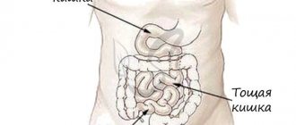
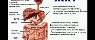
The intestine is the largest organ in the human body. The large intestine has an area of about 200 square meters. m., and the length is up to 2 m. The small intestine is longer (6-8 m). It is in this organ that the absorption of nutrients from food and medications occurs.
In addition, the intestines are a channel for cleaning the human body from waste products (feces). When the cleansing function is disrupted and the intestines become clogged, various pathologies occur. What to do if the intestines are clogged, and how to determine the pathology based on the symptoms.
Causes of intestinal pathology
If your intestines are clogged, it’s not easy to feel healthy
Where do the malfunctions in the work of this most important organ come from? Its activities are influenced by both external and internal factors. Preservatives, heavy metals, nitrates and pesticides that enter the body along with food and water are not completely eliminated, settling in the large intestine.
Improper, irregular nutrition also plays a big role. It’s not for nothing that the postulate “We are what we eat” exists. A person adds problems to himself by eating on the go, not chewing food properly, and abusing alcohol and medications. The causes of a clogged, slagged intestine are:
- High calorie foods
- Products that have undergone long-term heat treatment, canned food
- Lack of water in the body
- Low physical activity
- Stress
The listed reasons include the lack of a developed bowel movement regimen. As you know, going to the toilet should occur at the same time, preferably in the morning or evening. If a conscious habit of defecation at a certain time is not developed, feces begin to stagnate in the bends and folds of the intestine, gradually thickening and turning into fecal stones.
At the same time, the nerve endings of the intestines stop responding to the pressure of “waste” and the urge to go to the toilet occurs less and less often. Thus, a vicious circle arises: improper nutrition and diet provoke intestinal blockage, which in turn does not give signals for regular bowel movements, atony occurs, aggravating the blockage even more.
The weight of stones in the intestines can reach, according to various expert estimates, up to 3-5 kilograms, and this happens to both overweight and thin people.
Why cleanse your intestines?
Gastrointestinal upset is a huge problem for any person. After all, if the intestines are not cleaned and emptied in time, a number of diseases may arise.
Food is poorly digested, and energy goes to no one knows where. And the person feels bad - lethargic and overwhelmed. Therefore, you need to cleanse the body.
Of course, you need to cleanse all organs, but you need to start with the intestines. At the same time, there is an important rule: if the disease is obvious, and even more so in an advanced form, then you need to consult a doctor, because folk recipes can only be used prophylactically or in combination with the main treatment.
MICROBIAL FLORA OF THE INTESTINE AND DYSBACTERIOSIS
This lecture demonstrates the importance of intestinal microbal flora and it's modification in the disease for normal functioning of human's organism. The author stresses, that disbacteriosis is rather a condition than a desease observed in many intestinal and other gastro-intestinal tract pathologies. Clinical features of smaller and larger intestines disbacteriosis, as well as diagnostic patterns and treatment methods are presented.
A.I. Parfenov - Dr. honey. Sciences, prof., head Department of Small Intestine Pathology, Central Research Institute of Gastroenterology, Moscow AI Parfenov, MD, prof., Head Department of Small Bowel Pathology, Central Research Institute of Gastroenterology, Moscow
IN
In recent decades, great progress has been made in the study of microorganisms inhabiting the human intestine [1, 2]. The doctrine of intestinal dysbiosis has been formed [3–5]. In clinical practice, this bacteriological concept is often mistakenly identified with an independent nosological form.
Normal intestinal microbial flora and its role in human physiology
Normal microbial flora of the small intestine.
In the jejunum of healthy people, the environment can be sterile, although streptococci, staphylococci, lactic acid bacilli, other gram-positive aerobic bacteria and fungi are more often found in the upper sections.
The total number of bacteria in the jejunum does not exceed 104–105 per 1 ml of intestinal contents on an empty stomach. In the distal ileum, the number of microbes increases to 107–108, and anaerobic bacteria also appear [2]. Maintaining the normal ecology of the small intestine is ensured by the low pH of gastric juice, propulsive peristalsis, and efficient intestinal digestion and absorption [4]. Normal function of the ileocecal sphincter prevents reflux of colon contents into the small intestine. Normal microbial flora of the colon.
All colon microbes are divided into three groups: the main (bifidobacteria and bacteroides), constituting 70% of all bacteria, accompanying (lactic acid and E. coli, enterococci) and residual (staphylococci, fungi, Proteus).
Physiological role of intestinal microflora.
Microbial flora is necessary for the life of the macroorganism. Escherichia coli, enterococci, bifidobacteria and acidophilus bacilli have pronounced antagonistic properties. Under conditions of a normally functioning intestine, they are able to suppress the growth of microorganisms that are not characteristic of normal microflora. Therefore, germ-free animals and, possibly, patients with severe dysbiosis are more susceptible to infections [1]. The normal microflora of the colon takes part in the development of immunity. The breakdown of nutrients undigested in the small intestine in the large intestine is carried out by bacterial enzymes, resulting in the formation of various amines, phenols, organic acids and other compounds. Toxic substances formed in the intestines during microbial metabolism (cadaverine, histamine and other amines) are excreted in the urine and normally do not have a significant effect on the body. With dysbacteriosis, the level of amines in the blood can increase and be one of the reasons for the deterioration of the condition. Some of the amines (for example, histamine and serotonin) were included in regulatory systems during evolution. Under the influence of microflora enzymes, various processes of conversion of bile acids occur in the distal ileum: deconjugation, the conversion of primary bile acids synthesized in the liver into secondary bile acids. These diverse transformations are carried out by various microorganisms, each of which affects different stages of these processes. Under physiological conditions, 80 to 95% of bile acids are reabsorbed. The rest are excreted in feces in the form of bacterial metabolites. Their presence in the contents of the colon inhibits the absorption of water and prevents excessive dehydration of feces. Thus, the enzymatic activity of microflora contributes to the normal formation of feces. In the distal part of the intestine, microflora transforms bilirubin into stercobilin and urobilin. However, when microorganisms colonize the upper parts of the small intestine or when there is an excessive supply of bile acids and fatty acids in the large intestine, the enzymatic activity of the microflora becomes one of the important pathogenetic mechanisms of malabsorption in the small intestine and the development of diarrhea. Thus, microflora is an obligatory component of the “macroorganism - microorganisms” community.
Dysbacteriosis
The term “intestinal dysbiosis” means the appearance of a significant number of microbes in the small intestine and a change in the microbial composition of the large intestine. In the colon, the total number and properties of microorganisms change, their invasiveness and aggressiveness increase. The extreme degree of intestinal dysbiosis is the presence of gastrointestinal bacteria in the blood (bacteremia) or even the development of sepsis. Manifestations of dysbiosis in various combinations are found in almost all patients with chronic intestinal diseases, with some changes in nutrition and exposure to a number of environmental factors, and taking antibacterial drugs. Therefore, intestinal dysbiosis is a bacteriological concept, but in no case a diagnosis. Clinical manifestations of dysbiosis are largely determined by the localization of dysbiotic changes. Therefore, it is necessary to distinguish between dysbiosis of the small and large intestines. Dysbacteriosis of the small intestine (syndrome of increased bacterial contamination of the small intestine)
An increase in the number of bacteria in the small intestine may be associated with their excessive entry into the small intestine, favorable conditions for development and impaired propulsive function [2, 5, 7]. When bacterial contamination of the small intestine occurs, premature deconjugation of primary bile acids occurs. The resulting secondary bile acids and their salts cause diarrhea and are lost in large quantities in the feces. As a result, the development of gallstone disease is possible. Bacterial toxins, proteases, other metabolites, such as phenols, biogenic amines, bacteria can bind vitamin B12 [2]. Excessive microbial flora can lead to damage to the epithelium of the small intestine, since the metabolites of some microorganisms have a cytotoxic effect. There is a decrease in the height of the villi, deepening of the crypts, and with electron microscopy one can see the degeneration of microvilli, mitochondria and the endoplasmic reticulum [8]. With bacterial contamination, the secretion of water and electrolytes into the intestinal lumen increases, which causes diarrhea. The fat content in feces increases. The appearance of steatorrhea is associated with a decrease in conjugated bile acids in the intestinal lumen, which ensure emulsification of fats and activation of pancreatic lipase. With dysbiosis of the small intestine, the absorption of fat-soluble vitamins A, D and K is impaired.
Colon dysbiosis
The composition of the microflora of the colon can change under the influence of various factors and adverse effects that weaken the body’s defense mechanisms (extreme climatic and geographical conditions, pollution of the biosphere with industrial waste, various chemicals, infectious diseases, diseases of the digestive system, malnutrition, ionizing radiation). Iatrogenic factors play an important role in the development of colon dysbiosis: the use of antibiotics and sulfonamides, immunosuppressants, steroid hormones, radiotherapy, and surgical interventions. Antibacterial drugs significantly suppress not only pathogenic microbial flora, but also the growth of normal microflora in the colon. As a result, microbes that come from outside or endogenous species that are resistant to drugs (staphylococci, Proteus, yeast, enterococci, Pseudomonas aeruginosa) multiply. In most cases, the disturbed ecology of the colon gradually recovers on its own and does not require treatment [1]. In weakened patients, especially those with impaired immunity, self-healing of the intestinal ecology does not occur and clinical symptoms of dysbiosis appear.
Clinical features of dysbiosis
Clinical manifestations of excessive growth of microorganisms in the small intestine may be completely absent, be one of the pathogenetic factors of chronic recurrent diarrhea, and in some patients lead to severe diarrhea with steatorrhea, malabsorption syndrome and B12 deficiency anemia. With severe bacterial contamination syndrome observed in small intestinal diverticulosis, B12-deficiency anemia can be combined with peripheral neuropathy due to degenerative changes in the dorsal horns of the spinal cord. It should be emphasized that clinical symptoms associated with excessive growth of microorganisms in the small intestine and deviations in eubiosis of the large intestine are observed very rarely in clinical practice and are of more theoretical than practical importance. Severe malabsorption develops very rarely in patients with severe symptoms of stasis in the small intestine with partial intestinal obstruction and after surgical operations on the stomach and intestines. Particularly dangerous is pseudomembranous colitis, which is caused by toxins secreted by Pseudomonas aeruginosa Clostridium difficile. This anaerobic microorganism multiplies when the normal intestinal microbial flora is suppressed during treatment with broad-spectrum antibiotics. The main symptom of pseudomembranous colitis is profuse watery diarrhea, the onset of which was preceded by the prescription of antibiotics. Then cramping pain in the abdomen appears, body temperature rises, and leukocytosis increases in the blood. Very rarely, a fulminant course of pseudomembranous colitis, reminiscent of cholera, can be observed. Dehydration develops within a few hours and ends in death. V.N. Krasnogolovets distinguishes latent, common (with bacteremia) and common, occurring with generalization, infections (sepsis, septicopyemia) [3].
Methods for diagnosing dysbiosis
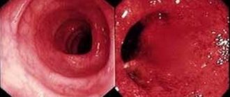
To diagnose dysbiosis of the small intestine, direct and indirect methods are used. Direct involves inoculating duodenal and jejunal contents obtained using a sterile probe. Excessive growth of bacteria is diagnosed if the number of bacteria exceeds 105/ml or microorganisms found in the large intestine (enterobacteriaceae, bacteroides, clostridia, etc.) are determined in it. It is known that in the process of metabolism of carbohydrates by the microbial flora of the colon, a large amount of gases, including hydrogen, are formed. This fact was the basis for the creation of a hydrogen test based on the determination of hydrogen in exhaled air. The hydrogen content in exhaled air is determined using gas chromatography or an electrochemical method [9]. The hydrogen test can be used to provide an approximate idea of the degree of bacterial contamination of the small intestine. This indicator is directly dependent on the concentration of hydrogen in exhaled air on an empty stomach. In patients with intestinal diseases occurring with chronic recurrent diarrhea and bacterial contamination of the small intestine, the concentration of hydrogen in exhaled air significantly exceeds 15 ppm. Lactulose loading is also used. Normally, lactulose is not broken down in the small intestine and is metabolized by the microbial flora of the colon. As a result, the amount of hydrogen in the exhaled air increases. With bacterial contamination of the small intestine, the “peak” appears much earlier. The most common bacteriological signs of colon dysbiosis are the absence of the main bacterial symbionts of bifidobacteria and a decrease in the number of lactic acid bacilli. The total number of microorganisms is often increased due to concomitant proliferation (Escherichia coli, enterococci, clostridia) or the appearance of residual (staphylococci, yeast-like fungi, Proteus) microflora. In addition to a change in the total number of microorganisms and a violation of the normal ratio between individual representatives of the intestinal microbial cenosis, the expression of dysbiosis can also be a change in properties with the appearance of pathological signs in individual bacterial symbionts. Hemolyzing flora, E. coli with weakly expressed enzymatic properties, enteropathogenic E. coli, etc. are detected. No specific features in the clinical course of the disease depending on one or another manifestation of dysbacteriosis in the colon have been established. It can be noted that patients with chronic intestinal diseases are more often infected with acute intestinal infections compared to healthy people, probably due to a decrease in the antagonistic properties of normal intestinal microflora and, above all, the frequent absence of bifidobacteria in them. The diagnosis of pseudomembranous colitis is established on the basis of bacteriological examination of stool and determination of Cl in them. difficile. The endoscopic picture is characterized by the presence of plaque-like, ribbon-like and solid “membranes”, soft but tightly fused to the mucous membrane. The changes are most pronounced in the distal colon and rectum.
Treatment of intestinal dysbiosis
Treatment of dysbiosis should be comprehensive. It includes: 1) elimination of excess bacterial contamination of the small intestine; 2) restoration of normal microbial flora of the colon; 3) improvement of intestinal digestion and absorption, 4) restoration of impaired intestinal motility; 5) stimulating the body's reactivity.
Antibacterial drugs
Antibacterial drugs are necessary primarily to suppress the excessive growth of microbial flora in the small intestine [10]. For this purpose, antibiotics from the group of tetracyclines, penicillins, cephalosporins, quinolones (ofloxacin) and metronidazole are usually used [11]. Broad-spectrum antibiotics significantly disrupt eubiosis in the colon. Therefore, they should be used only for diseases with impaired absorption and intestinal motility, in which, as a rule, a pronounced growth of microbial flora develops in the lumen of the small intestine. Antibiotics should be given orally in normal doses for 7–10 days. For diseases accompanied by colon dysbiosis, you should choose drugs that have minimal effect on the symbiont microbial flora and suppress the growth of Proteus, staphylococci, yeast fungi and other aggressive strains of microbes. These include antiseptics: intetrix, ersefuril, nitroxoline, furazolidone, etc. For severe forms of staphylococcal dysbiosis, antibiotics are used: ofloxacin, metronidazole, as well as co-trimoxazole, nevigramon. Antibacterial drugs are prescribed for 10–14 days. If fungi appear in the stool or intestinal juice, the use of nystatin or levorin is indicated. In the event of the development of pseudomembranous colitis, first of all, the antibiotic that caused the disease is discontinued. Prescribe vancomycin 125 mg orally 4 times a day; if necessary, the dose can be increased to 500 mg 4 times a day. Treatment is continued for 7–10 days. Metronidazole at a dose of 500 mg orally 2 times a day is also effective. In addition, bacitracin 25,000 IU orally 4 times a day is used. Bacitracin is almost not absorbed, and therefore a higher concentration of the drug can be created in the colon. In case of dehydration, adequate infusion therapy is used to correct the water and electrolyte balance. Cholestyramine (questran, vazazan) is used to bind the toxin.
Bacterial preparations
Bacterial preparations are used for diseases accompanied by colon dysbiosis. They can be prescribed without prior antibiotic therapy or after it. Bifidumbacterin [12], bificol, lactobacterin, bactisubtil [13], Linex [14], enterol [15] and other drugs have proven themselves well. The course of treatment should last 1–2 months. Recently, there have also been reports of the possibility of eliminating colon dysbiosis with various nutritional supplements [16–18]. A.L. Vertkin et al. [19] noted the normalization of the microbial flora in patients with colon dysbiosis after a course of treatment with the “NK” biococktail, consisting of biologically active extracts of vegetables, herbs and propolis, fermented with E. coli M-17. G.P. Minina et al. [20] reported the possibility of eliminating intestinal dysbiosis in children using the drug Nutrikon, containing bran, milk powder, bifidobacteria and medicinal herbs.
Products of microbial metabolism
Another possible way to eliminate dysbiosis is to influence the pathogenic microbial flora with metabolic products of normal microorganisms. Hilak-forte meets these requirements [13, 21], 1 ml of which corresponds to the biosynthetic active substances of 100 billion normal microorganisms. Hilak is prescribed 60 drops 3 times a day for up to 4 weeks in combination with antibacterial drugs or after their use. The drug is recommended for use in all forms of dysbacteriosis, both in combination with antibacterial drugs and as monotherapy.
Digestive enzymes and regulators of intestinal motility
A properly selected diet and enzyme preparations help improve digestion. For intestinal diseases accompanied by diarrhea, dietary nutrition should help restore impaired peristalsis and reduce the secretion of water and electrolytes into the intestinal lumen. The set of products must correspond in composition and quantity of nutrients to the enzymatic capabilities of the pathologically altered small intestine. The diet should be mechanically and chemically gentle, contain an increased amount of protein, and exclude refractory fats and foods to which tolerance is reduced. Diet No. 4b almost completely meets these requirements. In patients with disorders of cavity digestion of pancreatogenic origin, pancreatic enzymes have a good therapeutic effect. These include Creon, pancitrate, etc. For the treatment of steatorrhea of hepatogenic origin, drugs containing bile components (panzinorm, digestal, festal, enzistal, etc.) can be recommended. In case of gastrogenic insufficiency of digestion, it is advisable to use panzinorm containing hydrochloric acid and pepsin. To reduce flatulence, usually observed with dysbacteriosis, combination preparations have been created containing, in addition to enzymes, dimethicone (pancreoflat and zymoplex). In order to improve the absorption function, Essentiale, Legalon or Carsil are prescribed, which have a stabilizing effect on the cell membranes of the intestinal epithelium. Loperamide and trimebutine contribute to the restoration of impaired intestinal propulsive function.
Stimulants of body reactivity
To increase the reactivity of the body in weakened patients, it is advisable to use taktivin, thymalin, thymogen, immunal, immunofan and other immunostimulating agents. The course of treatment should last an average of 4 weeks. Vitamins are prescribed at the same time.
Prevention of dysbacteriosis
Primary prevention of dysbiosis, given the numerous causes of its occurrence, is a very difficult task. Its solution is associated with general preventive problems: improving the environment, rational nutrition, improving well-being and other numerous factors of the external and internal environment. Secondary prevention involves the rational use of antibiotics and other medications that disrupt eubiosis, timely and optimal treatment of diseases of the digestive system, accompanied by a violation of microbiocenosis.
Literature:
1. O.V. Chakhava, E.M. Gorskaya, S.Z. Ruban. Microbiological and immunological foundations of gnotobiology. M.-Medicine. - 1982;160. 2. Farthing MJG. Bacterial Overgrowth of the Small Intestine., Gastroenterology, Misiewicz G (ed) // New York, 1993;2:45. 3. V.N. Krasnogolovets, Intestinal dysbiosis. M. 1989;208. 4. A.M. Ugolev. The evolution of digestion and the principles of the evolution of functions. 1985; L. Science.-543. 5. Saltzman JR, Russell RM. Nutritional consequences of intestinal bacterial overgrowth.//Compr.Ther. - 1994;20:523–30. 6. Fried M., Siegrist H., Frei R., et al. Duodenal bacterial overgrowth during treatment in outpatients with omeprazole. // Gut. 1994;35(1):23-6. 7. Saltzman JR; Kowdley KV; Pedrosa MC; et al: Bacterial overgrowth without clinical malabsorption in elderly hypochlorhydric subjects.: //: Gastroenterology. 1994;106(3):615–23. 8. Haboubi NY, Lee GS, Montgomery RD Duodenal mucosal morphometry of elderly patients with small intestinal bacterial overgrowth: response to antibiotic treatment.// Age-Ageing; 1991;20:29–32. 9. Loginov A.S., Lorie N.Yu., Parfenov A.I. and others. Diagnostic capabilities of determining hydrogen in exhaled air - a new functional test for intestinal diseases. // Ter.arch. 1991;2:74-6. 10. Corazza GR, Sorge M, Strocchi A, et al. Non-absorbable antibiotics and small bowel bacterial overgrowth.// Ital. J. Gastroenterol. 1992;24:4-9. 11. Levin MS. Medical management of diseases of the small intestine.// Curr. Opin Gastroenterol. 1992;8:224-31. 12. A.Yu. Zorin, Yu.V. Kozminykh, R.R. Kildiyarova. Comparative clinical and microbiological aspects of the use of dry and liquid bifidobacterins in children with dysbiosis. On Sat. “Man and Medicine” M.-1998;275. 13. Parfenov A.I., Kaloev Yu.K., Safonova S.A. and others. Intestinal dysbiosis. Moscow medical journal.-1998;1:12-7. 14. N.P. Brashkina, G.A. Samsygina, G.D. Pershina et al. Linex in the treatment of intestinal dysbiosis in young children. .// On Sat. “Man and Medicine” M.-1998;257. 15. Matveev V.A., Yesenina O.I., Karpova E.Yu. and others. Enterol in the treatment of intestinal dysbiosis.// In collection. “Man and Medicine” M.-1998;135. 16. Gnoevykh V.V., Vize-Khripunova M.A. Floradophilus and intestinal dysbiosis. .//Rus. magazine gastroenterology, hepatology, coloproctology. 1997;5:97. 17. Podkorytov Yu.A. The use of biologically active food additives for intestinal dysbiosis. .// On Sat. “Man and Medicine” M.-1998;396. 18. Starostin B.D. Laminolact in the treatment of diseases accompanied by intestinal dysbiosis. .//Rus. magazine gastroenterology, hepatology, coloproctology. 1997;5:111–2. 19. Vertkin A.L., Artamonov V.E., Bagaturia I.F. and others. Experience of using the bio-cocktail “NK” for the correction of colon dysbiosis in patients with peptic ulcer disease.// In collection. “Man and Medicine” M. 1998;40. 20. Minina G.P., Lupan I.N., Nosal M.F. Experience of clinical use of the nutritional supplement Nutrikon for disorders of intestinal microecology in children. .// On Sat. “Man and Medicine” M.-1998;299. 21. Babushkin N.V. Use of the drug “Hilak-forte” in the complex treatment of intestinal dysbiosis. .//Rus. magazine gastroenterology, hepatology, coloproctology. -1997;5:96–7.